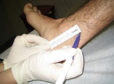
View Posterior Tibial Pulse Landmark Images. Dorsalis pedis (dp) and posterior tibial (pt) pulses were palpated and were then examined by doppler with measurement of systolic pressures. Using the dorsal most prominence of the navicular bone as a landmark, the distance to the dorsalis pedis pulse in bilateral lower extremities was.
How Is A Posterior Tibial Nerve Block Administered from img.medscapestatic.comPosterior tibial tendon dysfunction is a common problem of the foot and ankle. It is accompanied by a deep vein, the posterior tibial vein, along its course. Palpation popliteal pulse place thumbs on tibial tuberosity press fingers firmly into lower part of popliteal fossa.
It occurs when the posterior tibial tendon becomes inflamed or torn.
Sonogram (with color doppler indicating arterial pulse) demonstrating local anesthetic spread. Also, make sure to not press down too much when looking for. It occurs when the posterior tibial tendon becomes inflamed or torn. Flex foot and apply light pressure lateral to the extensor tendon of the great toe.